Through the organs of vision, a person receives most of the information. This is why it is so important to take care of your eyes and vision from childhood. Often, the weakening of visual functions occurs unnoticed, which raises the question of regularly visiting an ophthalmologist and checking visual acuity.
In this article
- Why is it important to have your eyes checked?
- How is visual acuity checked?
- Sivtsev table
- How is vision checked using the Sivtsev table?
- Optotypes of Pole
- Golovin table
- Snellen chart
- Siemens star
- Duochrome test
- Amsler grid
- How is vision checked in children?
- Ways to test children's vision at home
- Determination of visual acuity in preschool children
- How to measure vision yourself?
- How often should you have your eyes checked?
- The first symptoms of visual impairment
Why is it important to have your eyes checked?
More and more people are facing vision problems. Neither children nor adults are immune from them. This is due to poor nutrition, poor environment, constant use of computers and mobile gadgets and other factors. Very often, people do not notice that their vision has deteriorated or simply do not pay attention to the first symptoms, attributing their appearance to fatigue and eye fatigue. This may cause the refractive error or visual pathology to progress. This is especially dangerous in childhood, when identifying an eye disease and stopping its development is most important.
The future full life of the child will largely depend on this. To avoid complications, it is necessary to regularly visit the ophthalmologist’s office, regardless of whether the first symptoms of vision loss appear or not.
How is visual acuity checked?
Visual acuity is the ability of the eye that allows you to see two objects or two points located at a certain distance from each other, separately. This function of the visual apparatus is one of the most important; it depends on the width of the pupil, refraction, transparency of the lens, cornea and vitreous body, the condition of the retina, optic nerve, as well as on age and other factors.
Visual acuity is determined in the ophthalmologist's office using computer equipment and tables. With the help of instruments, the doctor examines the fundus of the eye, the condition of the retina and the eye as a whole, and calculates various parameters that will be required to select correction means - glasses and contact lenses. In addition, tests and other procedures may be required to determine the causes of deterioration in visual function. Visual acuity is determined using special tables, the most famous of which is Sivtsev’s table. It is familiar to every person from school age. There are other methods. Let's find out what their features are and how to check your vision yourself, without turning to a specialist.
Sivtsev table
Perhaps everyone remembers the poster with the large black letters ШБ located at the very top. This is a table for determining visual acuity, invented by the scientist, ophthalmologist Dmitry Aleksandrovich Sivtsev. It is a group of printed letters - optotypes. There are only seven of them: B, I, K, Sh, Y, M, N. They are written in 12 lines in different orders. Starting from the top line, optotypes decrease in size. To the right of the lines, the value corresponding to visual acuity is indicated. On the poster it is indicated by the Latin letter V, and it is expressed in a conventional unit (not to be confused with diopters), with which a person can distinguish the letter from a distance of five meters (0.1, 0.2, 0.3 and so on). To the left of the letters is another value - the distance from which a person with good eyesight should be able to read the letter freely. This parameter is designated by the letter D.
What is color blindness?
Color blindness is a pathology of the visual system in which a person is unable to reliably distinguish colors. Most often, the anomaly occurs among males.
In this article
- What is color blindness?
- Features of the color perception process
- Myths and truth about color blindness
- Testing for color blindness: what is the Rabkin table?
- Why are drivers tested for color blindness?
- How is color blindness tested?
- Can color blindness be cured?
The disease occurs due to disorders associated with the pigments of the retina. It is often transmitted from mother to son in the womb (due to disturbances at the gene level, in the X chromosome). But there are many cases where, during a color blindness test, it turns out that the disease is not hereditary in nature, but is classified as acquired. This is possible with various types of injuries to the organs of vision, in which the integrity of the retina is disrupted, which subsequently disrupts the functions of the cones. For the first time, the chemist J. Dalton, who suffered from this disease, spoke about color blindness. In his works, the first records of color blindness were made in 1794.
How is vision checked using the Sivtsev table?
The table hangs on the wall and is illuminated by two fluorescent lamps. Lighting should be 700 lux. The lower edge of the lamp is located at a height of 120 cm from the floor. Each eye is checked separately. The subject sits on a chair five meters from the table and covers one eye with a shutter. You need to keep your head strictly straight. The doctor points to the optotype with a pointer, and within two to three seconds the person being tested must name the letter.
Visual acuity is defined as complete if a person correctly named all the signs, and incomplete when errors are made, but their number is limited - no more than one in lines from the first to sixth and no more than two in lines from seven to ten.
If the result obtained is below 0.1, then the patient is nearsighted (myopia), if above 0.1 - farsighted (hyperopia). Normal refraction of the eye is called emmetropia, that is, for such a person the point of clear vision is at a distance of five meters or more. When an object is placed at a distance of less than five meters, the rays are collected in parallel on the retina. In this regard, exactly five meters are considered the optimal distance for visometry.
If the patient is unable to distinguish optotypes even in the very top line, which with ideal vision can be seen from 50 meters, he is asked to move half a meter closer to the table (or closer depending on the need). Then visual acuity is calculated using the formula V = d / D. D is the distance for a person with good vision, and d is the actual distance from which the patient sees the letters in the table. If the person being tested does not see the letters of the first row (visual acuity is lower than 0.1), Pole optotypes are used.
Diagnostics using the Rabkin method
In most cases, ophthalmologists determine the degree of color sensitivity impairment, as well as its features, using Rabkin tables. These are 27 pages of cards on which the design is applied in the form of colored spots and dots of equal brightness but different shades. Depending on what kind of visual impairment a person suffers from, he can distinguish individual images, and sees some pictures in monochrome.
Diagnostics using the Rabkin method
How the test is taken
In order for the results to provide objective information, the test is carried out under the following conditions:
- normal general health and mood;
- positioning the picture at the same level as the eyes of the person being tested;
- looking at the picture for no more than 10 seconds.
Otherwise, the results will be unreliable.
Table for studying Rabkin's color perception
Decoding tables
In order to convert the test results into a diagnosis, it is important to know what deviations are revealed by the special vision of each picture. It should be borne in mind that only an ophthalmologist can accurately decipher the results, and the test will be as informative and accurate as possible when it is performed using paper rather than electronic media, because the settings of a particular computer can change the true test colors
So:
card 1. On it is the number “96”. The table does not carry a special diagnostic load, as it is intended more for explanation and familiarization with the test;
Card 1
card 2. Here you can distinguish a square and a triangle. Those who do not see this are malingerers;
Card 2
card 3. On it is the number “9”. A colorblind person will say that this is a “5”;
Card 3
card 4. In this figure, a person with normal color vision sees a triangle, and with deviations, a circle;
Card 4
card 5. The number “13” is visible. Those suffering from color blindness will claim that they see “6”;
Card 5
card 6. On it there is a circle and a triangular figure. A person who has poor color vision will not see them;
Card 6
card 7. The number “9” should be seen by both people with normal and problematic color perception. Do not see? Malingerers;
Card 7
card 8. The number “5” on it is visible only to healthy people;
Card 8
card 9. For those who do not distinguish shades of red, it will seem like “8” or “6” in the picture. And only people with normal color vision will see nine;
Card 9
card 10. Anyone who sees “68”, as well as “66” or “69” in this picture, has problems with the perception of colors. A healthy person will find “136” here;
Card 10
- card 11. The number “14” should be read here by both people with normal vision and those with visual impairments;
- card 12. The number “12” is visible here, but those suffering from a lack of perception of the color red will not see this;
Card 12
card 13. The picture shows a circle and a triangle. Those who have problems with the color green will only find a triangle. If red is not perceived, only a circle will be visible;
Card 13
card 14. On it are the numbers “3”, “6” and “0”. If you are unable to distinguish green, “1” and “6” will be visible. And if there are problems with red – “1”, “0” and “6”;
Card 14
card 15. Only healthy people can distinguish between a circle and a triangle with a square. Those who have trouble identifying colors will offer other answers;
Card 15
picture 16. On it is the number “96”, which can be easily read by a person with normal vision. If there are difficulties with the red spectrum, then only nine will be visible. When there is a problem with green, a six will be noticeable;
Card 16
picture 17. The triangle and circle depicted here will only be seen by healthy people. People with impaired color perception recognize only one of the figures;
Card 17
picture 18. In this picture, a healthy person will see squares of different colors arranged in horizontal and vertical rows. If colors are misperceived, assumptions arise about the monochrome nature of some rows or columns;
Card 18
picture 19. On it is the number “95”. A person with a color anomaly will only see “5”;
Card 19
picture 20. The person being tested, who has a healthy perception of colors, will describe a circle and a triangle here. A colorblind person will not see them.
Card 20
As for pictures No. 21 to No. 27, they repeat those described above.
Optotypes of Pole
Polyak's optotypes are a method for determining visual acuity, named after the Soviet ophthalmologist Boris Lvovich Polyak. He created his method specifically for military medical and medical-social examination, during which disability or suitability for military service is identified. Optotypes are sticks, strokes, rings depicted on the poster, which are located at a fairly close distance from the patient’s eyes. The width of the gaps between the strokes, as well as the thickness of the lines, determines visual acuity in the range from 0.04 to 0.09.
How is color blindness tested?
The visual organs are tested for color sensitivity at an appointment with an ophthalmologist during a routine examination of the patient. You can also take the test using Rabkin’s tables and get an answer online. However, you should not completely trust the results of taking the test remotely, especially if potential signs of color vision anomalies are identified. The correctness of testing is influenced by many factors (lighting, monitor settings, etc.), so for specifics you will need to visit an ophthalmologist.
Golovin table
It was created by oculist Sergei Selivanovich Golovin. It is practically no different from the table for testing vision proposed by Sivtsev, and they are used, as a rule, together, but it uses Landolt rings as optotypes - black circles broken on one side. The rings on the poster are located similarly to the optotypes in Sivtsev’s table. Golovin’s method is more reliable, because remembering rings and deceiving an ophthalmologist is much more difficult than in the case of letters.
Snellen chart
To determine visual acuity, a technique developed by the Dutch ophthalmologist Hermann Snellen is also used. The table was created in 1862 and is still considered one of the most reliable and accurate. It consists of capital Latin letters. It has 11 lines. Letters decrease in size from top to bottom. The largest optotypes can be distinguished by a person with good vision from a distance of 60 meters. Also, normal vision allows you to see signs located below the first large line from distances of 36, 24, 18, 12, 9, 6, 5 meters.
During the examination, the table is located at a distance of 6 meters from the patient. He closes one eye and reads optotypes with the other. Visual acuity is indicated by the lowest row, which was recognized accurately. The normal indicator is 6/6. In this case, the bottom lines from six meters differ. If the patient normally sees only the lines that are located above the row visible from 12 meters by a person without refractive errors, then his visual acuity is 6/12. The doctor will determine the values in negative and positive diopters.
This table has Latin notations, in addition, it shows distances in feet, so it is used mainly in Western countries.
Siemens star
This is another technique that is not a table, but a star consisting of 54 black rays on a white background. The diameter of the star is 10 cm. The rays converge from the periphery to the center of the circle. A person without eye refractive errors from five meters observes how the rays merge exactly in the middle, they begin to overlap each other. This happens when 2.5 cm remains to the center of the star. At a distance of more than five meters from the picture, the rays gather into a solid mass of gray color.
In the presence of refractive errors, the rays seem to merge with the background and overlap each other. However, closer to the center they can again be clearly visible. The star begins to appear as its own negative, that is, the white background becomes black, and the black rays become white. If you have good eyesight, then you can observe this effect by placing the picture close in front of your eyes.
This technique allows you to determine not only myopia and hypermetropia, but also astigmatism. With this pathology, the outer boundary of the rays does not form a circle, but an ellipse or an even more complex figure.
Vision for license category B (BE, B1)
The simplest restrictions apply for drivers of passenger cars:
12. Visual acuity is below 0.6 in the best eye and below 0.2 in the worst eye with tolerable correction with 2 eyes open, regardless of the type of correction (spectacles, contact, surgical), the degree and type of ametropia or eye length.
Let's figure out how to use this point in practice:
- Have your visual acuity measured by an ophthalmologist. You will receive 2 numbers (one for each eye).
- Choose the larger one of the two numbers. If it is equal to or greater than 0.6, then everything is in order, a certificate will be issued. You don't have to watch the next steps.
- If the visual acuity of the “better” eye is 0.5 or less, then move on to the second number. If the second digit is 0.2 or more, then a certificate will be issued.
- If the visual acuity of the “worst” eye is 0.1 or less, then a certificate will not be issued.
Let's look at a few examples to better understand in what cases a certificate will be issued.
Left eye 1.0; right eye 1.0. The visual acuity of the better eye is 1.0, it is more than 0.6, that is, a certificate will be issued.
Left eye 0.8; right eye 0.5. The visual acuity of the better eye is 0.8, it is more than 0.6, that is, a certificate will be issued.
Left eye 0.6; the right eye is missing. The visual acuity of the better eye is 0.6, it is equal to 0.6, that is, a certificate will be issued. For category B, the absence of one eye is not a reason for not issuing a certificate.
Left eye 0.2; right eye 0.5. The visual acuity of the worse eye is 0.2, it is equal to 0.2, that is, a certificate will be issued.
Left eye 0.2; right eye 0.2. The visual acuity of the worse eye is 0.2, it is equal to 0.2, that is, a certificate will be issued.
Left eye 0.1; right eye 0.5. Visual acuity of the better eye is 0.5, it is less than 0.6. The visual acuity of the worse eye is 0.1, it is less than 0.2, that is, a certificate will not be issued.
Thus, a driver whose vision is (0.5; 0.1) or worse will not be able to obtain a medical certificate.
If at least one eye has a visual acuity of 0.6 or better, or two eyes have a visual acuity of 0.2 or better, then the certificate will be issued without any problems.
Duochrome test
The duochrome test is used to determine myopia and farsightedness. The table is a rectangle divided into two halves. One of them is red (left), the other (right) is painted green. The letters are located exactly in the middle, the same as in Sivtsev’s table. The purpose of the test is to determine in which color field the patient distinguishes optotypes better. If he sees letters better on a red background, then he suffers from myopia, if on a green background, he suffers from hypermetropia. If the symbols are clearly visible in both fields, then the vision is excellent, that is, we are talking about emmetropia.
This test must be taken with glasses if there is already a diagnosed visual pathology. After checking, it is necessary to adjust the optical power of the lenses.
Amsler grid
This test is needed to check the central field of vision and allows you to identify such ophthalmological diseases as macular degeneration of the retina, scotomas (dark spots in the eye), metamorphopsia (distortion of objects, their shapes, colors, sizes).
The grid (also called a grid) is a large black square with numerous small black squares inside on a white background. There is a large black dot in the center of the reticle. The test is carried out as follows:
- The Amsler grid is located at the patient’s eye level at a distance of 30 cm;
- the patient sits on a chair with his back straight;
- one eye is covered with the palm of your hand (you cannot put pressure on it, otherwise the results will be erroneous);
- the other eye looks at the central point for 5 seconds;
- then you need to approach the grid by 10 cm and look at the point for another 5 seconds;
- takes the starting position at a distance of 30 cm from the net;
- the process is repeated with the second eye.
In the absence of ocular pathologies, all lines and angles of the grid are straight. When the lines are distorted and bent, we can talk about disturbances in the retina. One of the conditions of the test is that it must be taken in the optics that the patient uses in everyday life.
The doctor decides which table to check your vision. It all depends on the specific case, the patient’s complaints and the progress of the examination.
The essence of testing using Rabkin tables
Testing is carried out by an ophthalmologist using special test tables. They allow you to establish the quality of color perception and identify pathology or simulation.
To check, Rabkin’s polychromatic tables are used, which are shown in a certain sequence for 5 seconds.
Moreover, the structure of the tables is such that it allows one to detect and diagnose not only the presence of pathology, but also to find out its origin (congenital or acquired).
Rabkin's table is 27 basic test pictures that allow you to establish the presence of congenital or acquired visual pathology in a short time, as well as 20 additional tables to clarify the diagnosis; they help verify the correctness of the diagnosis.
How to learn a table for vision?
Is it possible to memorize a table and deceive the doctor? Some people ask this question and search the Internet for techniques for quickly memorizing tables. When a person wants to get a driver's license of a certain category, and he has vision problems, he may try to resort to such deception. However, the specialist immediately determines the presence and absence of refractive errors by focusing and the patient’s eyes. In addition, in addition to tables, instruments are also used that cannot be deceived. Therefore, asking such a question is pointless. Do not try to deceive the doctor, so as not to get into an awkward situation.
Reference images
To identify color blindness, a set of tables is used, which consists of 27 images.
Below are pictures and explanations that will help you assess your health.
1
The number 96 is shown here; it can be determined by both healthy people and sick people. This table was created primarily to clearly show a person the principle of testing and help him orient himself.
2
A healthy person should see geometric shapes here. A square and a triangle are presented. This picture makes it possible to identify malingerers, since the figures are easily distinguished between sick and healthy people.
The figure shows the number 9. A person with progressive color vision impairment will only be able to see 5 here.
4
The patient will see a triangle without deviations. If you can only see a circle, then you have health problems.
5
Healthy people will be able to see 13 of the circles, but pathological changes in the ocular apparatus will allow them to see only 6.
6
This table is one of the most difficult. Healthy people will look at the triangle and the circle in the picture. If you have the disease, it will be difficult to notice any figures at all.
7
With full color perception, you can see the number 9. The vast majority of sick people are also able to see this number.
8
In this case, it is necessary to discern the number 5. It will be quite difficult for people with color blindness to complete this task, but still some cope with it.
9
Even those people who have problems recognizing the green spectrum can easily identify the number 9. If a person has problems with the perception of the color red and its shades, then it may seem that the picture shows 8 or 6.
10
If there are no problems with vision, then seeing the number 136 will not be a problem. With pathological changes, a person may see the number 68,69 or 66.
11
14 can be seen by almost all patients who undergo this test. Colorblind people see this image well, so if determining the numbers is difficult, then it is worth undergoing a full examination of the eye apparatus.
12
Considering the number 12 will be difficult for people who do not distinguish the red spectrum. In all other cases there should be no problems.
In this case, geometric shapes are located. The circle and triangle can only be seen by people who are not colorblind.
14
Partial blindness that does not recognize the red or green spectrum will prevent patients from viewing all the numbers. Healthy people will identify numbers 6,3 and 0.
15
This image shows 3 geometric shapes at once. Even a healthy person cannot always see the square at the bottom and sometimes only sees a circle and a triangle. If there is poor perception of the red spectrum, then a person will recognize 2 triangles.
16
The doctor gives an assessment of health depending on how many numbers the person was able to see. If he sees the number 96, then most likely there are no problems in the eye apparatus. When only one number is detected, color blindness occurs.
The clear delineation of the two halves of the image allows almost everyone to detect the triangle and the circle.
18
A healthy person will see in this table that the horizontal rows are made in the same color, and the vertical ones in different colors. If red or green spectrum blindness is present, the results will be different.
The number 25 will be easy to see for people with good eyesight. If there is an anomaly, only the five will be visible.
20
The triangle and circle here can only be seen by those patients who do not have problems with color perception. If there is at least partial blindness in the green or red spectrum, then it will be impossible to see the figures.
21
The numbers in this picture can be seen by a healthy and a sick person. A small number of people are unable to see the number 9 because they have difficulty distinguishing the green spectrum.
It is very difficult for colorblind people to see the number 5 in this picture, but almost everyone copes with this task.
Healthy people will see single-color vertical lines in this table. And they will note that the horizontal cubes are made in different colors. With color blindness, the patient will give the opposite readings.
A person must see the number 2 in this image, otherwise he has a disease.
Here is exactly the same number as in the previous picture.
26
The inverted triangle will be clearly visible if there are no problems with the perception of the green spectrum. If there is a violation, the patient will only be able to see the circle.
How is vision checked in children?
All of the above vision testing tables can only be used if the patient knows the alphabet. How to test visual acuity in preschoolers? This is very important, since it is necessary to identify refractive errors as early as possible in order to stop the progression of the pathology. In children, the eyeball is just developing and you need to carefully monitor all changes to prevent the occurrence of serious diseases, for example, strabismus.
Babies undergo their first vision test as soon as they are born. It is performed by a neonatologist. He examines the baby's eyes without using any instruments. However, such a test can only give a superficial opinion about the condition of the child's eyes. It allows you to identify obvious congenital anomalies. If any are detected, the neonatologist gives a referral to an ophthalmologist.
The first scheduled vision test is carried out at 3 months. Next - at 6 and 12 months after birth. Here the ophthalmologist already uses special instruments during the examination.
Why are drivers tested for color blindness?
Colorblindness in drivers is dangerous both for the person driving the car and for those around him. With complete color blindness, the ability to fully monitor the situation on the road is reduced. The driver, even with normal refraction, will have difficulty reading road signs, get confused by traffic lights, etc.
Therefore, checking for color blindness is a mandatory condition for a future driver to pass a medical examination, without which a license will not be issued in Russia. Thus, a person who is colorblind is legally prohibited from driving. But in real life, many people with this pathology continue to drive. Especially with a partial violation of color perception, which is not critical and does not interfere with control of the situation on the road.
Ways to test children's vision at home
Parents can also assess a child’s visual abilities at home. They can observe the baby's behavior and the reaction of his eyes when showing him various objects. Babies' pupils must respond to light, and at two months they begin to fixate their eyes on objects. By six months, children recognize familiar objects. At one year old, babies can see small objects from a distance of one meter. By about two years, this distance will be 2.5 meters. If you notice any deviations from the norm, immediately make an appointment with a specialist.
Visual acuity test
Each of us has already had an eye exam at an ophthalmologist's office, but what if you want to monitor your visual acuity every day? Is it possible to test your eyesight at home? Yes, it is possible, and completely free. There are two options.
First option:
Download the vision measurement chart and print it out. We offer you 3 types of tables:
Table for measuring visual acuity Sivtsev download
Table for measuring visual acuity Golovin download
Table for measuring vision for children Orlova download
Instructions “how to measure vision using downloaded and printed tables”
1.Print the table on A4 sheet.
2. Position the sheet so that the 10th line (the line above the gray stripe) is at eye level. The distance between the eyes and the table is 2.5 meters.
3. Turn on good lighting in the room, the table should be illuminated evenly.
4. Cover one eye with your palm or something opaque (do not close it under any circumstances).
5. Mark the line that you see with each eye separately (to the right of each line is the value V, this is the value of your vision. For example, you see the 4th line from the top - that means your vision is 0.4).
Second option:
Online visual acuity test. We offer a unique algorithm for measuring visual acuity online, in which you do not need to know the resolution, diagonal size and proportions of your monitor. In this case, you will get a result that is as close as possible to the real one (however, visual acuity can be measured most accurately in an ophthalmology office) by synchronizing the dimensions of the monitor with the dimensions of the table. Also, a very important factor for the reliability of measuring visual acuity is the absence of distractions, therefore, when checking vision online on our website, you will see a table on a white background, without banners or advertising. All you need for an online vision test is to have a banknote of the appropriate denomination or a ruler with you.
Instructions “how to measure vision online on the website infoglaza.ru”
1.Attach a sample (bill, card or ruler) to the monitor and mark it as shown in the figure. Select the sample you took (what you measured). Set the desired distance from the monitor. Select a table to measure visual acuity. Click on the “start vision test” button
2.Move away from the monitor so that there is the distance you set between your eyes and the monitor.
3. Cover one eye with your palm or something opaque (do not close it under any circumstances).
4. Mark the line that you see with each eye separately (to the right of each line is the value V, this is the value of your vision. For example, you see the 4th line from the top - that means your vision is 0.4)
Determination of visual acuity in preschool children
Between the ages of three and seven, a child can already speak, but may not yet know the alphabet. Then Orlova’s table comes to the rescue. It resembles the tables of Golovin and Sivtsev, but instead of rings and letters, it contains pictures (mushrooms, stars, trees, animals) that the child is able to name.
The principle of the examination is the same. The preschooler is at a distance of five meters from the poster. Looking at it with one open eye, he begins to name the images shown by the ophthalmologist. Since children get tired faster than adults, the ophthalmologist begins to ask about the pictures in the top rows and selects only one from each row. If the child is not able to give the correct answer, then he is shown another image in the same row. This is done to make sure that the little patient really does not see the object well, and does not just not know its name.
How to measure vision yourself?
Can a person use an ophthalmologist's chart to test their vision at home? In principle, nothing prevents this. However, in the ophthalmologist’s office all conditions have been created for a full examination. Moreover, only a doctor can make the most accurate measurements. Testing visual acuity at home is only needed to roughly find out about the presence of vision problems. If you independently determine visual impairment, you need to go to the clinic and undergo a detailed examination.
So, if you still decide to carry out visometry at home, then you can make a Sivtsev table yourself. You will need three white matte A4 sheets, a printer, a black marker, tape and glue. You find the necessary files on the Internet and print them. If the contrast is not very high, then it can be increased using a marker or felt-tip pen. Three printed sheets are connected with tape and hung on a door, wall or cabinet.
When calculating the height, make sure that the tenth line is at your eye level. A 40 W lamp is suitable for uniform illumination of the table. It needs to be directed to the poster. Next, you close one eye and look at the table from a distance of five meters. Since it is difficult to measure the distance and lighting level as accurately as possible at home, you will get an approximate result.
The importance of color vision testing
Color blindness does not have a particular impact on the quality of life, as evidenced by the fact that people most often learn about the presence of this congenital pathology in adulthood. However, in some cases it can become an obstacle to engaging in one or another type of activity.
A number of highly specialized professions require the absence of color blindness. Thus, for drivers this special test is mandatory before obtaining a driver’s license; young men pass it at the military registration and enlistment office. Color vision testing is carried out for sailors, pilots and a number of other professions.
Can I use a computer to test my vision?
All of the tests listed can be taken online. There are many programs that offer their own methods and exercises, but you can also use tables used by ophthalmologists. To ensure that the results are as close to reality as possible, you should follow a number of rules when taking tests on a computer:
- you need to be at a distance of 30-50 cm from the monitor;
- the monitor should be at eye level in front of you so that its surface does not cast glare;
- testing is carried out only when you feel well (fatigue can distort the results);
- the table should be clear and contrasting;
- When taking the test, you should not squint or strain your eyes.
Today, vision is checked both with the help of conventional paper tables and computer programs, and with the help of a variety of transparent instruments. The optotypes are not placed on paper, but on milk glass (opaque frosted glass). The light source is behind this glass. He displays images and letters on it. Ophthalmologists also use a collimator method for determining visual acuity. Special devices create an optical system with optotypes that are located directly in front of the eye. This method is necessary when it is not possible to arrange a distance of five meters between the table and the patient. Collimator devices are used for mass examinations. Also in clinics you can see how ophthalmologists work with projectors that provide good illumination, clear and contrasting images without distortion and glare.
Diagnostic methods
There are many types of color blindness. The easiest one is the inability to distinguish shades of color. For example, dark pink is perceived as red, while lighter tones are seen as white.
Rabkin tables
The most famous and popular diagnosis is using Rabkin tables. They depict many colorful circles of different shapes and sizes. All these circles are of the same brightness, and among them numbers or geometric shapes stand out in a different color.
A person with normal vision can almost easily distinguish them from the rest. If there are violations, these encrypted figures will be indistinguishable to him.
Testing
It happens simply - signs are placed in front of the subject in good, uniform lighting, the patient examines them for a few seconds and says what he sees. In this case, the person should be in a relaxed state and feel good. 7-10 seconds are allotted for viewing one picture; the pictures are located at a distance of 1 meter from the eyes at the same level with them.
These tables also contain trap figures. For example, a person with normal color vision sees 9, and a colorblind person sees 5, or a circle. Only colorblind people with a certain type of colorblindness see them; the doctor, using his tables, will be able to determine which pigment is missing on the retina.
Several pictures in the set were created to identify malingerers, those who for some reason want to deceive the signs when passing the test. These plates depict pictures that are clearly and clearly visible with normal vision and with any form of color blindness. The doctor can easily determine which color the patient cannot distinguish.
The circles on the tables are selected in such a way as to check all possible deviations. And the color combinations in them are not random. However, when taking the test on a healthy person, errors are possible - the result may depend on the computer screen itself and the lighting.
There are 48 plates in the set, 27 are used for general diagnostics. The rest are needed for a more in-depth analysis. They are necessarily included during inspections of professional drivers, machinists, and pilots.
The main Rabkin tables can be purchased, the rest remain for more detailed analysis from a doctor. It is useless for a colorblind person to pretend to have healthy vision; the doctor will decipher it anyway. It is also useless to pretend to be colorblind during military commissions - the remaining plates will still show color perception, it is impossible to learn them.
If these tables show severe color vision impairment, for example, red and green look the same, the person will be denied a license altogether. The same thing will happen if colors in the blue-yellow range are not distinguished.
Testing for color blindness using the Rabkin table is used all over the world; it gives the most reliable result, the type and degree of pathology. And control pictures will clarify the diagnosis.
Yustova's technique
This technique also consists of cards and is designed to identify which color disorder the patient has.
The subject is offered four sets of cards, each of them identifying the pathology of one primary color.
- The first set reveals disturbances in the perception of red and its shades,
- second - green spectrum
- third - blue
- fourth - black and white text
The tables are special pictures with “torn” squares, which depict a stylized letter C, with a break in one of the sides of the square. The subject must determine the location of the rupture. There are 12 tables of the test, each table shows several pictures with such an image. All cells form one tone, the test is constructed in such a way that the possibility of “learning” it is excluded.
The test is also good because it is easy to conduct with children who do not yet know numbers and geometric shapes.
This test is also popular among ophthalmologists around the world. It is also used in diagnosing acquired color blindness in adults after various injuries, diseases and taking certain medications.
Ishhara Test
The test is somewhat reminiscent of Rabkin's tables. In it, the subject is also offered tables consisting of many circles of different colors and sizes. But in it a person must no longer distinguish numbers or letters, but pictures and simple images.
This test is used mainly to diagnose color blindness in children; they like such pictures and willingly guess the encrypted images. These pictures can also be offered to those who have “learned” Rabkin’s tables for “passing a medical examination.” If color blindness is present, a person will not be able to distinguish the desired image.
Some other color blindness tests work in much the same way. These are the color schemes of Stilling, Schaaf, and some others. Their principle is approximately the same.
How often should you have your eyes checked?
Many people wonder how often they need to have their eyes checked if nothing is bothering them. It depends on age and other factors. Newborns are examined on the first day, at three months, at six months and at one year. Schoolchildren undergo annual medical examinations, during which their vision is also checked. It is also advisable for adults to visit an ophthalmologist once a year. People over 60 years of age can do this more often. Much depends on specific conditions. So, it is imperative to check the condition of the visual organs during pregnancy in order to avoid retinal detachment during childbirth.
If there are problems with refraction and correction means are used - glasses or contact lenses, then vision is checked every 6 months. You may need lens dioptre adjustments.
There are groups of people who need to visit an ophthalmologist regularly. These include those who:
- faced various eye diseases, eye injuries;
- underwent eye surgery;
- has a predisposition to ophthalmological diseases;
- treated with hormonal drugs;
- suffers from vascular and nervous system diseases;
- planning a pregnancy.
It is also recommended for hypertensive patients and diabetics to regularly visit the ophthalmologist's office.
Penalty for driving without means of correction when there are restrictions
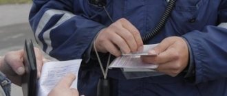
If the driver has successfully passed the medical examination wearing glasses or contact lenses, he can drive a vehicle only when using vision correction devices. A special GCL mark is made about this in the license on the reverse side of the license.
If a driver drives a car without vision correction, such actions are equivalent to driving a car without a license. In such a case, the provisions of Part 1 of Art. 12.7 of the Code of Administrative Offenses of the Russian Federation, according to which he will be fined in the amount of 5 to 15 thousand rubles.
Share on social networks:
More useful on the topic
How to properly start a manual vehicle: step-by-step instructions for beginners
23.05.2021
How to properly adjust the side and interior mirrors of a car yourself
08.04.2021
The first symptoms of visual impairment
There are many reasons why vision deteriorates. They can be infectious and non-infectious, congenital and acquired. If you feel that your vision has become worse, then you should not think about the reasons, but it is better to immediately go to the doctor. Only he, after conducting a detailed examination, will establish the causes, make a diagnosis and prescribe treatment or write a prescription for correction agents. However, there are several signs that may indicate a deterioration in visual function. They are usually noticed quickly. Therefore, it is important not to simply ignore them, but to take appropriate measures.
There are three clear signs of vision loss:
- Inability to see objects that were previously quite easy to see. For example, you cannot see letters when writing or reading; they become blurry. At the same time, if you squint your eyes, they are clearly visible again.
- It is not possible to see inscriptions on store windows, signs on houses and other texts at a great distance.
- Objects and objects lose their brightness, become dull, blurry, unclear.
If these symptoms develop rapidly, then measures to stabilize vision and stop the progression of the pathology should be taken immediately.
Today there are many factors that have a negative impact on the eyes. But medicine is also developing rapidly. You can check your vision at home or in any clinic. Moreover, various companies offer a huge selection of correction products (glasses and contact lenses of all types for every buyer’s taste). You just need to take more care of your health: eat right, spend time in the fresh air more often, lead an active lifestyle, maintain eye hygiene, perform visual exercises if your work involves eye strain, and regularly check with an ophthalmologist.
Myths and truth about color blindness
Color blindness certainly affects a person's quality of life. For example, for potential drivers, this factor may become the main reason for refusal to issue a license when passing a mandatory medical commission. Moreover, patients who are unable to distinguish colors are somewhat limited in their choice of profession.
There are many rumors associated with color blindness. Let's figure out which of them are true and which belong to the category of myths:
- People suffering from color blindness see the world around them in black and white and cannot distinguish green from red. This definition of color blindness is not entirely reliable, since there are several types of pathology, and with some of them people cannot distinguish only certain shades related to certain colors.
- Color blindness is a purely male pathology. This statement is also wrong. Indeed, the majority of people affected by the disease are men, but many cases of color blindness have been recorded in women (mostly these are girls to whom the disease was inherited, that is, there were already precedents for color blindness in the family).
- It is impossible to develop color blindness as an adult. As already noted, the anomaly can be both congenital and acquired. It may be preceded by various retinal injuries.
It is also interesting that with a normal visual examination it is impossible to determine color blindness in a person - the pathology is not visually reflected in the eyes. Therefore, special diagnostic methods were invented, in which pathology is diagnosed using tables and cards. The most popular way to determine or refute this anomaly is considered to be Rabkin’s cards and tables.